Arteriovenous Malformation (AVM): Understanding the Condition and Its Treatment
13 August, 2026
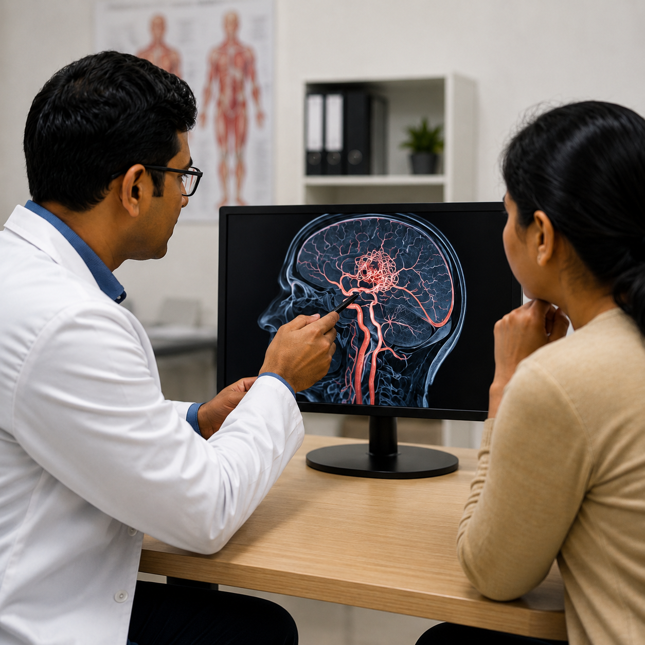
Arteriovenous malformation (AVM) is a rare condition where there is an abnormal formation connecting the arteries and veins. In a healthy person, the blood flows from arteries through capillaries into veins. However, in AVM, the normal sequence is bypassed, allowing the blood to flow directly from arteries to veins.
An AVM can occur in various regions of the body. However, brain arteriovenous malformations (brain AVMs) demand special care because of their ability to cause hemorrhage and neurological complications. An AVM can be asymptomatic in some people and result in headaches and seizures in others.
Thanks to improvements in diagnostic imaging technology and minimally invasive surgical techniques of
B. P. Poddar Cancer Hospital, AVMs are now being diagnosed and treated with several therapeutic options available for consideration.
What Happens in an Arteriovenous Malformation?
Typically, arteries conduct blood under high pressure, whereas veins receive blood from the arteries in a low-pressure state. The capillary bed between them acts to control blood circulation.
In an AVM, the presence of capillaries is not there, causing direct transfer of blood from arteries to veins. It leads to unusual blood flow and can also add to the stress in the vessels.
In certain situations, the blood vessels can be prone to rupture, thus leading to bleeding. Nevertheless, it must be noted that not all AVMs are associated with any complications.
Symptoms of Brain AVM
The symptoms of a brain AVM can vary depending on its location, size and whether it has caused bleeding.
Some patients may experience:
- Persistent or recurrent headaches
- Seizures
- Weakness or numbness
- Problems with speech or vision
- Difficulty with coordination or movement
- Changes in neurological function
In some cases, an AVM may remain silent and be discovered incidentally during brain imaging performed for another reason.
A sudden and severe headache, sudden weakness, difficulty speaking, loss of consciousness or other sudden neurological symptoms can indicate a medical emergency and require immediate medical attention.
Why Can an AVM Be Serious?
Among the issues related to brain AVM is the occurrence of brain hemorrhage, in the case where there is any rupturing of the abnormal blood vessels.
The chances of developing the complication do not affect all patients equally. Things such as the site of the AVM, the AVM's size and configuration, and prior bleeds are taken into account when evaluating the AVM.
That is why AVM needs proper evaluation instead of generalized treatment methods.
How Is an AVM Diagnosed?
Accurate diagnosis is an important part of AVM management. Doctors use advanced imaging techniques to understand the location and structure of the abnormal blood vessels.
Depending on the patient's symptoms and clinical condition, investigations may include
MRI, CT scans and cerebral angiography.
Cerebral angiography is particularly useful for understanding the blood vessels supplying and draining the AVM. This detailed information helps specialists plan treatment and assess the relationship between the AVM and surrounding brain structures.
Treatment Options for AVM
Treatment for an arteriovenous malformation depends on several factors, including its size, location, blood-flow pattern, symptoms and the potential risks associated with treatment.
In some patients, careful observation with regular imaging may be appropriate. When active treatment is recommended, modern management may involve
endovascular embolization, microsurgery, stereotactic radiosurgery, or a combination of these approaches.
Endovascular Embolization
Endovascular embolization is a minimally invasive technique used to reduce blood flow through the abnormal vessels.
A thin catheter is guided through the blood vessels to reach the AVM. Specialized material can then be delivered through the catheter to block or reduce blood flow to selected vessels feeding the malformation.
Embolization may sometimes be used as an independent treatment in carefully selected cases, or as part of a planned treatment strategy before surgery or radiosurgery.
Microsurgical Treatment
For certain AVMs, particularly those that can be safely accessed,
microsurgical removal may be considered.
During the procedure, the abnormal vessels are carefully identified and separated from the surrounding healthy brain tissue before the AVM is removed.
Whether surgery is appropriate depends heavily on the AVM's location and its relationship with important areas of the brain. Detailed pre-operative imaging and specialist assessment are therefore essential.
Stereotactic Radiosurgery
Stereotactic radiosurgery is a non-invasive treatment that uses precisely focused radiation to target the abnormal blood vessels.
Unlike conventional surgery, it does not require an open surgical incision. Over time, the treated vessels undergo changes that can gradually lead to closure of the AVM.
This approach may be considered for selected AVMs, particularly when the malformation is small or located in an area where conventional surgery may carry greater risks.
When a Combination of Treatments May Be Needed
Some AVMs are complex and may not be best managed with a single treatment method.
In selected cases, specialists may use a
multimodal approach, combining techniques such as embolization and surgery or radiosurgery. For example, embolization may be performed first to reduce blood flow through specific parts of the AVM before another treatment is undertaken.
The objective is to achieve effective treatment while minimizing the potential risk to surrounding healthy brain tissue.
How Doctors Decide the Right Treatment
AVM treatment needs to be personalized. Before recommending a particular procedure, specialists may evaluate:
- The size and location of the AVM
- The arteries supplying the malformation
- The veins draining it
- Whether the AVM has previously bled
- The patient's symptoms
- The relationship of the AVM to important areas of the brain
- The potential risks and benefits of each treatment option
This detailed assessment helps the medical team determine whether observation, embolization, surgery, radiosurgery or a combination of treatments is most appropriate.
Conclusion
An
AVM requires precise diagnosis, careful planning and specialised treatment like SGRT. At
B. P. Poddar Hospital Cancer Unit, advanced technology like IGRT, SGRT from
Vision RT’s Align RT and multidisciplinary expertise come together to provide personalised care tailored to each patient’s condition.
The right evaluation is the first step towards the right treatment.


 NABH Accredited Hospital